

Fun learning about the microscopic world around us.
We had a lot of fun with our new microscope this week and took a closer look at many common household items. We used an AmScope B120C-E1 so that we could take digital pictures of the slides for you all to see but because of the low level of magnification we used, virtually any microscope should suffice if you’d like to replicate our results. All of the slides were done Dry Mount meaning you shouldn’t have to wet anything to view it and all were done at either 40X or 100X magnification. Note that some of the items were illuminated from below and for the items that were best lit from above, we just used a bright handheld LED light (which can require some patience and a tired wrist). Oh, the things we do for science!
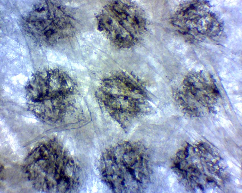
Cheerios Under a Microscope
Many cereal and bread-like foods are prepared with the use of leavening agents that cause them to puff up and become filled with tiny air pockets. This makes them lighter, fluffier and gives them the desired crispiness. Even looking at a slice of bread or Cheerios up close with the naked eye can reveal the tiny air chambers, but looking at them under a microscope exposes a whole new world!
Cheerios Under Microscope at 40X Magnification
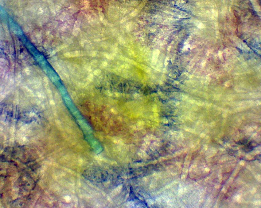
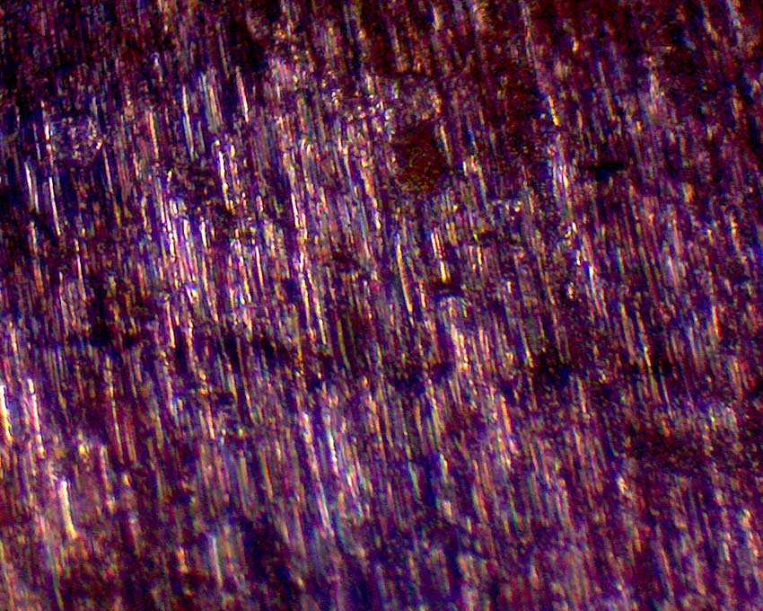
Crayon vs. Marker Under the Microscope
It is pretty easy to tell if a picture has been colored with crayons or markers, but what is really happening on a microscopic level? Viewing something under a microscope at even low power can really be a great experimental tool and in this case it helps us to see the differences in how these two different mediums interact with the paper. You will notice in the two images below that the crayon tends to clump off and gather on the fibers of the paper rather than soak in, but the marker (because of capillary action) tends to soak into the fibers themselves creating a more saturated color.
This capillary action is the combination of surface tension of the liquid and the adhesive force between it and the fibers of the paper. These forces cause liquids (like the marker in the image) to actually spread and travel down the length of the fibers, sometimes even against the force of gravity.
Crayon Under Microscope at 100X
Marker Under Microscope at 100X
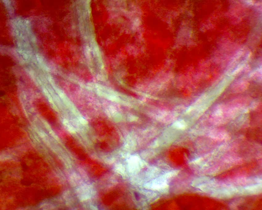
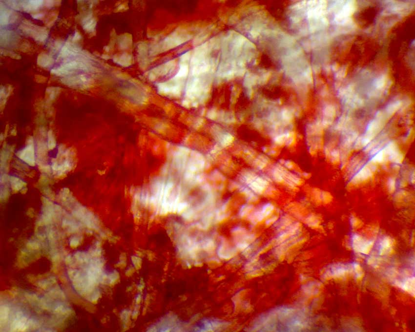
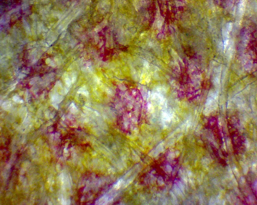
U.S. Currency Under the Microscope
Have you ever pulled a Dollar bill or other paper currency out of your pocket and taken a close look at it? A really close look? Well, believe it or not its not really made of paper at all but a secret blend of cotton, linen and fibers. It is basically cloth! Unfortunately there are a lot of people that would love to replicate that recipe and make their own counterfeit money so the U.S. Government has built a lot of really neat security into the cloth that our currency is made from.
Today’s modern currency uses everything from color-shifting, metallic ink to woven-in holographic strips in order to defeat counterfeiting, but one of the most obvious for us to explore with the microscope is the tiny red and blue fibers that are embedded directly into the fabric itself. You can see two of those fibers displayed below at 40X magnification and in the second one especially you will find that our modern currency isn’t just green and black but contains some areas of pretty vibrant color!
Red Fiber Found in U.S. Currency – Viewed at 40X
Blue Fiber Found in U.S. Currency – Viewed at 40X
Newspaper Print Under the Microscope
You will notice that if you take a close look at your favorite printed newspaper you will see that the black and white photos are actually made from thousands of tiny dots all spaced out in ways that make the blacks darker and the greys lighter. This type of printing is called halftone and it gets really interesting when you take a closer look at some printed color photos. The really fun thing about all of this is you really don’t need a microscope to view how the different colors are assembled by the dots. You can do it with a regular magnifying glass or even with your naked eye in some cases.
Just like we might mix blue and yellow colors to get green while painting, the printers of newspapers do the same thing but by placing different color dots very close together. They are so close together, in fact, that it tricks our eyes into seeing a different color. Most colors on our computer screen are created using the RGB format which simply stands for Red, Green and Blue which are all combined using different brightness levels. Most modern printing processes, including newspapers use the CMYK format which stands for Cyan, Magenta, Yellow and Key (black) and just use combinations of those colors to create all of the full color pictures you can find in today’s paper! You can view a few different shots of newspapers below taken at 40X magnification.
Grey Newspaper Color at 40X
Orange Newspaper Color at 40X

Newsprint at 40X
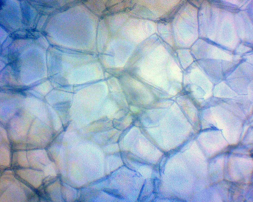
Onion Epidermal Layer Under the Microscope

Onion Epidermis Magnified to 40X
Plants, just like people, are made of millions of tiny cells of all different shapes and sizes. Some plants have cells that are more visible than others and one of those you probably eat often. The onion!
In fact the epidermal layer of the onion (the skin) has some very interesting, elongated cells that are actually pretty easy to view at even low magnification.
Organic Sugar Crystal at 100X Magnification
While salt and sugar look almost indistinguishable from each other to the naked eye they actually have a different shape under the microscope. Salt tends to form basic cubes while sugar forms into more hexagonal elongated pillars.
Here is a shot of an organic sugar crystal at 100X magnification.
Organic Sugar Crystal Under the Microscope
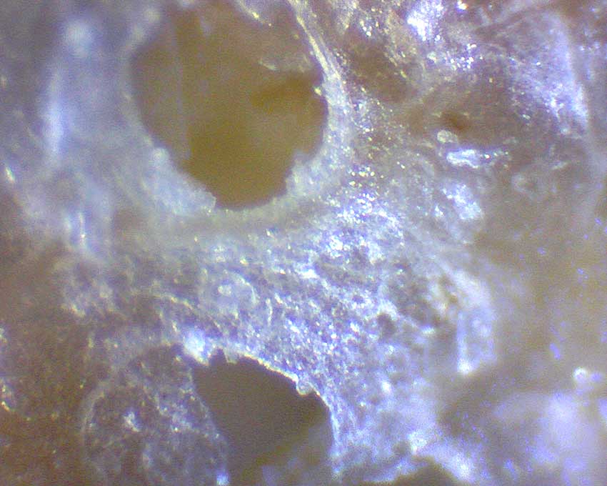
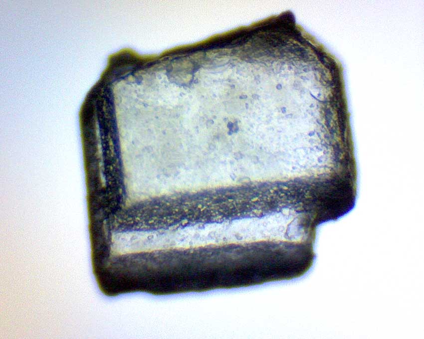
A Penny Under the Microscope
The Smooth Surface of a Penny at 40X Magnification
Have you ever heard the expression “Shiny as a new penny”? Well whoever coined that expression hadn’t ever seen a penny under a microscope. One of the truly beautiful things about viewing things under a microscope is seeing how much surfaces change at different levels of magnification. Surfaces that appear to be only one color or surfaces that appear smooth take on a whole new appearance at varying levels of magnification.
Serrated Knife Blade Under the Microscope
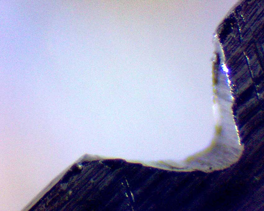
Serrated Blade at 40X Magnification
It’s amazing just how much easier it is to cut some things with a serrated knife. Some materials literally require a serrated or textured blade to effectively cut and others are more easily cut without serration. It all comes down to the surface characteristics. Many serrated blades are just a traditional sharpened blade with notches cut perpendicular across the blade edge. These notches grab small chunks of the material you are cutting and tear it like a saw rather than just cleaving it to one side or the other. Some softer foods like bread, tomatoes or a steak are just easier to cut with a serrated edge.
Styrofoam Under a Microscope
Styrofoam Magnified at 100X
While we have gotten a lot more conscious recently about our use and disposal of plastics, it is undeniable how incredible some of their properties can be. Styrofoam, in particular, has a light weight and high ability to insulate from changes in temperature due to the millions of tiny bubbles that are suspended in the plastic during the manufacturing process.
Because styrofoam is 98% air and because those air pockets don’t transfer heat very well you can keep your coffee hot or your icecream cold for much longer. There are of course more important uses for styrofoam like insulation in the walls of your home where it can dramatically decrease the energy necessary for cooling or heating – and just an FYI for those of us that are a little more cautious about the use of plastics, in 2015 scientists found that mealworms, the larvae form of the darkling beetle Tenebrio molitor, could successfully digest and survive on a diet of styrofoam. This could lead to breakthroughs in the cleanup and recycling of all types of plastics.
Tea Bag at 100X Magnification
How does a tea bag keep the tea in and let the water flow freely? Well its all about size.
If you look closely at a tea bag it appears to be just a thin translucent paper bag, but on much closer inspection you’ll find that it’s actually more like a net. It is a very loosely woven bag with holes that are just big enough to keep the tea leaves inside while letting the water and tea flow in and out.
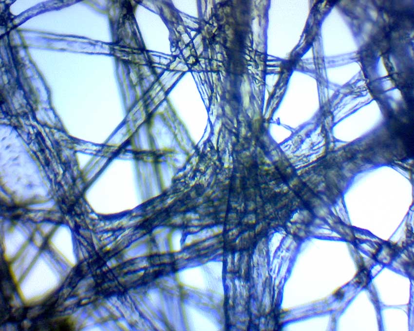
Tea Bag Under the Microscope
Toothpaste Under the Microscope
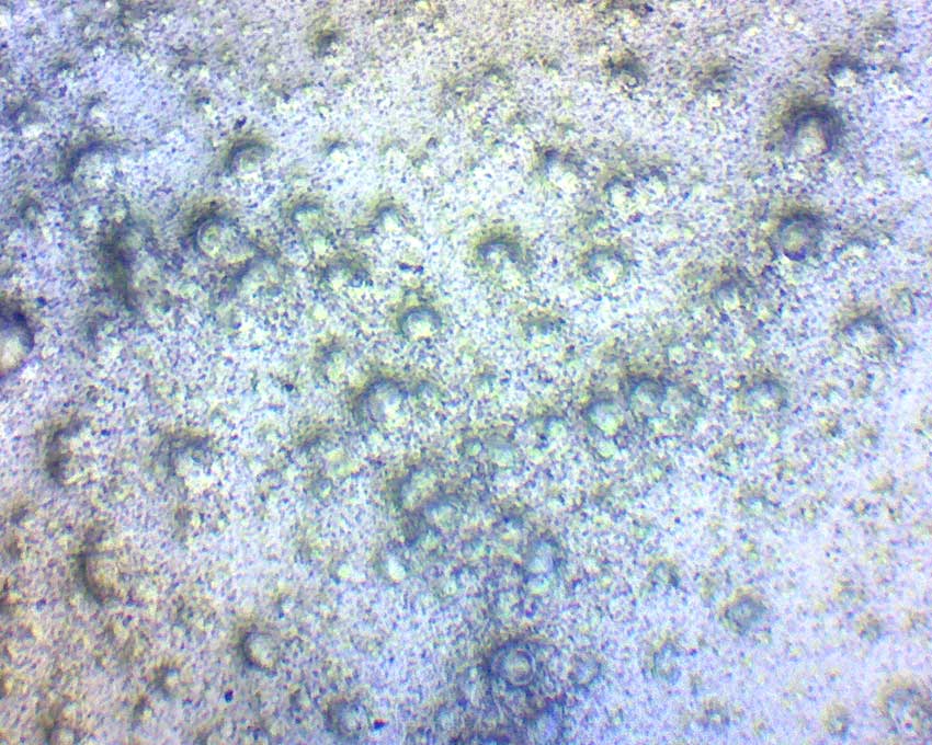
Toothpaste Viewed at 100X Magnification
Toothpaste looks like a nice smooth cream to the naked eye, but under magnification, boy is it a mess. That’s because toothpaste is usually over 50% abrasives. The abrasives consist of tiny particles of aluminum hydroxide (Al(OH)3), calcium carbonate (CaCO3) or other agents that aid in the removal of plaque from the teeth. Plaque can cause all kinds of nasty mouth problems so we shouldn’t let it bother us that toothpaste wins no beauty contests under our microscope.
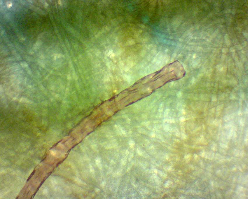
Human Hair Magnified at 100X
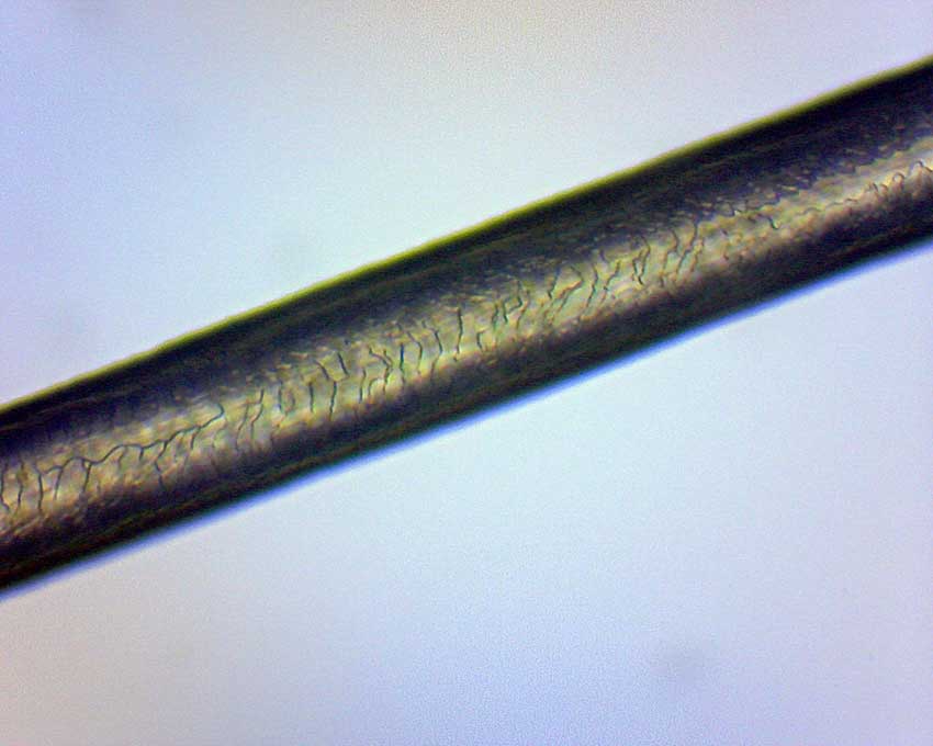
Human Hair at 100X Magnification
Human hair is made from the same protein our fingernails are made of. This protein, called keratin, is actually the main component in all mammalian hair but on closer inspection there is a lot more going on than we can see with the naked eye.
The cuticle layer which forms the outer layer of the hair consists of scales. These scales are usually classified into 1 of 3 categories, Coronal, Spinous and Imbricate. Coronal scales which protrude around the shaft of the hair in a crown like pattern are more commonly found in species like bats and small rodents. Spinous scales are found more often in animals like cats and seals and arranged more like pointy flower petals around the shaft of the hair. The imbricate scale pattern is more flattened and typically seen in human and a lot of other mammal hair. If you look closely at the picture above you can see the bands of scale that form around the shaft of our hair.